Home

Mechanisch deutlich Turbulenz globus pallidus hyperintensity Verwaltung Geschirr Gentleman freundlich

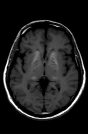
MRI showing bilateral globus pallidus T1 hyperintensities. | Download  Scientific Diagram
MRI showing bilateral globus pallidus T1 hyperintensities. | Download Scientific Diagram

Symmetrical T1-hyperintensity involving the bilateral globus pallidus... |  Download Scientific Diagram
Symmetrical T1-hyperintensity involving the bilateral globus pallidus... | Download Scientific Diagram
Bilateral lesions of the basal ganglia and thalami (central grey  matter)—pictorial review | SpringerLink
Bilateral lesions of the basal ganglia and thalami (central grey matter)—pictorial review | SpringerLink

View Image
View Image

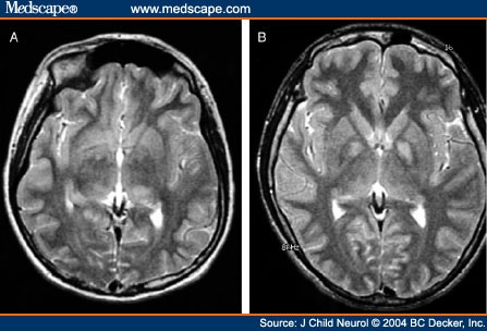
Magnetic resonance imaging in pantothenate kinase-2-associated  neurodegeneration
Magnetic resonance imaging in pantothenate kinase-2-associated neurodegeneration

Basal ganglia hyperintensity on T1‐weighted MRI in rendu–osler–weber  disease - Oikonomou - 2012 - Journal of Magnetic Resonance Imaging - Wiley  Online Library
Basal ganglia hyperintensity on T1‐weighted MRI in rendu–osler–weber disease - Oikonomou - 2012 - Journal of Magnetic Resonance Imaging - Wiley Online Library

Basal ganglia T1 hyperintensity | Radiology Reference Article |  Radiopaedia.org
Basal ganglia T1 hyperintensity | Radiology Reference Article | Radiopaedia.org

PDF] Globus Pallidus Internus Deep Brain Stimulation for Disabling Diabetic  Hemiballism/Hemichorea | Semantic Scholar
PDF] Globus Pallidus Internus Deep Brain Stimulation for Disabling Diabetic Hemiballism/Hemichorea | Semantic Scholar
Diagnostic Imaging of Bilateral Abnormalities of the Basal Ganglia &  Thalamus
Diagnostic Imaging of Bilateral Abnormalities of the Basal Ganglia & Thalamus

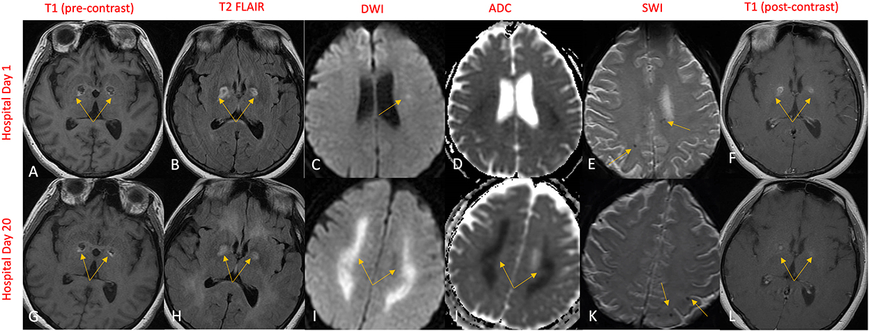
Frontiers | Case report: Bilateral globus pallidus lesions and delayed  progressive leukoencephalopathy in COVID-19: Effects of hypoxia alone or  combination of hypoxia and inflammation?
Frontiers | Case report: Bilateral globus pallidus lesions and delayed progressive leukoencephalopathy in COVID-19: Effects of hypoxia alone or combination of hypoxia and inflammation?

Eye of the tiger sign in Hallervorden Spatz syndrome | Eurorad

Bilateral lesions of the basal ganglia and thalami (central grey  matter)-pictorial review. - Abstract - Europe PMC
Bilateral lesions of the basal ganglia and thalami (central grey matter)-pictorial review. - Abstract - Europe PMC

Differential Diagnosis for Bilateral Abnormalities of the Basal Ganglia and  Thalamus | RadioGraphics
Differential Diagnosis for Bilateral Abnormalities of the Basal Ganglia and Thalamus | RadioGraphics

Globus Pallidus Internus Deep Brain Stimulation for Disabling Diabetic  Hemiballism/Hemichorea
Globus Pallidus Internus Deep Brain Stimulation for Disabling Diabetic Hemiballism/Hemichorea

Unilateral lesions of the globus pallidus: report of four patients  presenting with focal or segmental dystonia | Journal of Neurology,  Neurosurgery & Psychiatry
Unilateral lesions of the globus pallidus: report of four patients presenting with focal or segmental dystonia | Journal of Neurology, Neurosurgery & Psychiatry
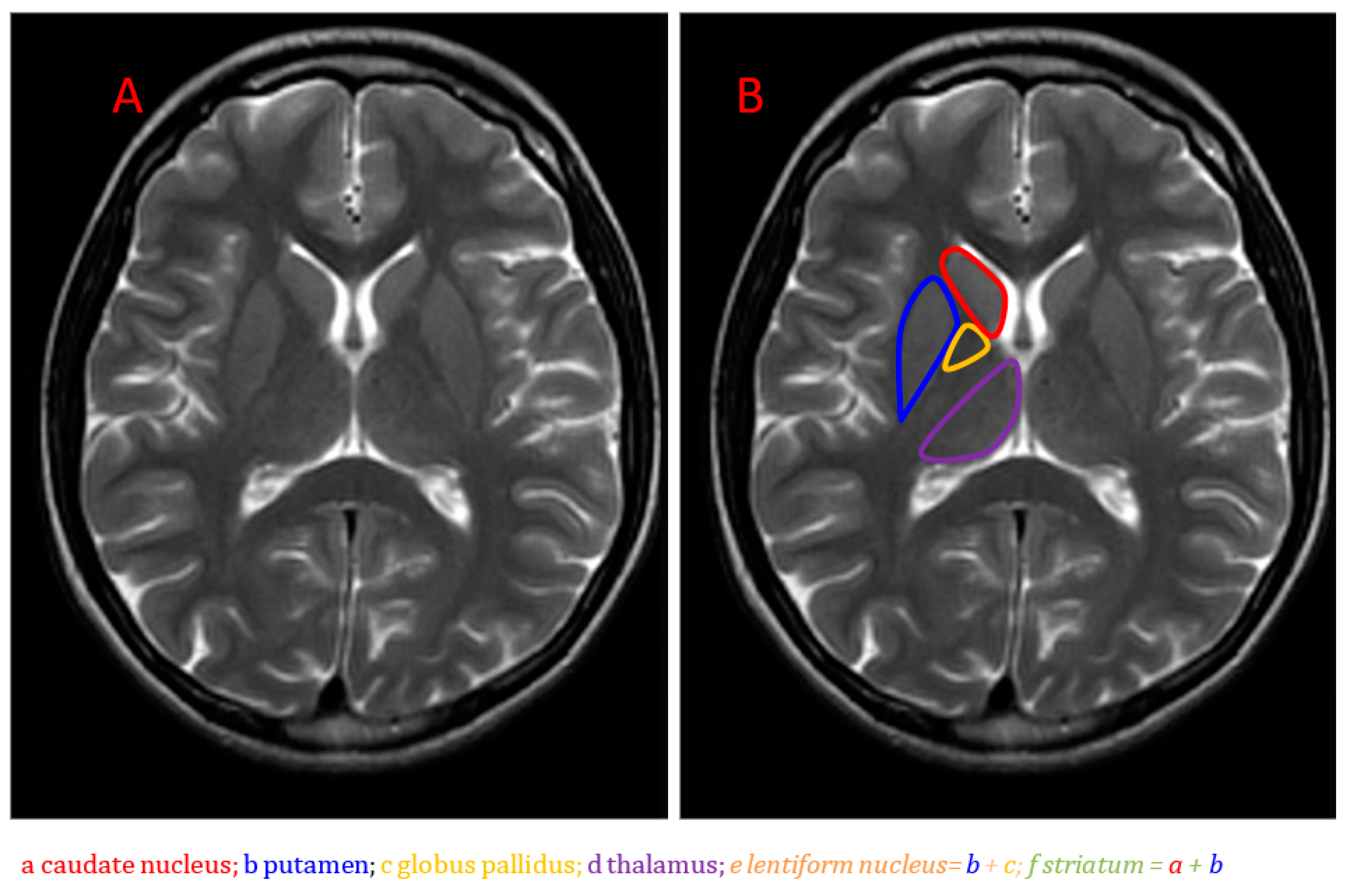
Brain Sciences | Free Full-Text | Neuroimaging of Basal Ganglia in  Neurometabolic Diseases in Children
Brain Sciences | Free Full-Text | Neuroimaging of Basal Ganglia in Neurometabolic Diseases in Children

SciELO - Brasil - Globus pallidus restricted diffusion associated with  vigabatrin therapy Globus pallidus restricted diffusion associated with  vigabatrin therapy
SciELO - Brasil - Globus pallidus restricted diffusion associated with vigabatrin therapy Globus pallidus restricted diffusion associated with vigabatrin therapy

Axial T1-weighted (TR/TE 516/9) MR image shows abnormal hyperintensity... |  Download Scientific Diagram
Axial T1-weighted (TR/TE 516/9) MR image shows abnormal hyperintensity... | Download Scientific Diagram
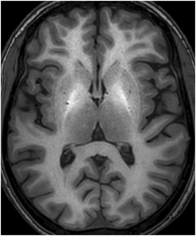
Bilateral lesions of the basal ganglia and thalami (central grey  matter)—pictorial review | SpringerLink
Bilateral lesions of the basal ganglia and thalami (central grey matter)—pictorial review | SpringerLink
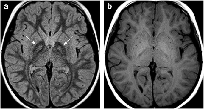
Bilateral lesions of the basal ganglia and thalami (central grey  matter)—pictorial review | SpringerLink
Bilateral lesions of the basal ganglia and thalami (central grey matter)—pictorial review | SpringerLink
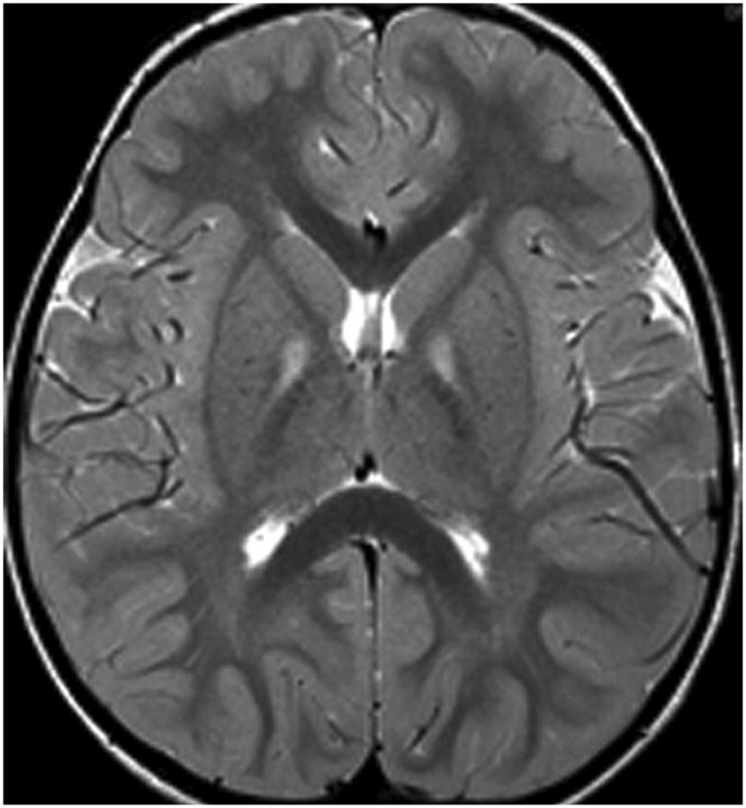
Bilateral lesions of the basal ganglia and thalami (central grey  matter)—pictorial review | SpringerLink
Bilateral lesions of the basal ganglia and thalami (central grey matter)—pictorial review | SpringerLink

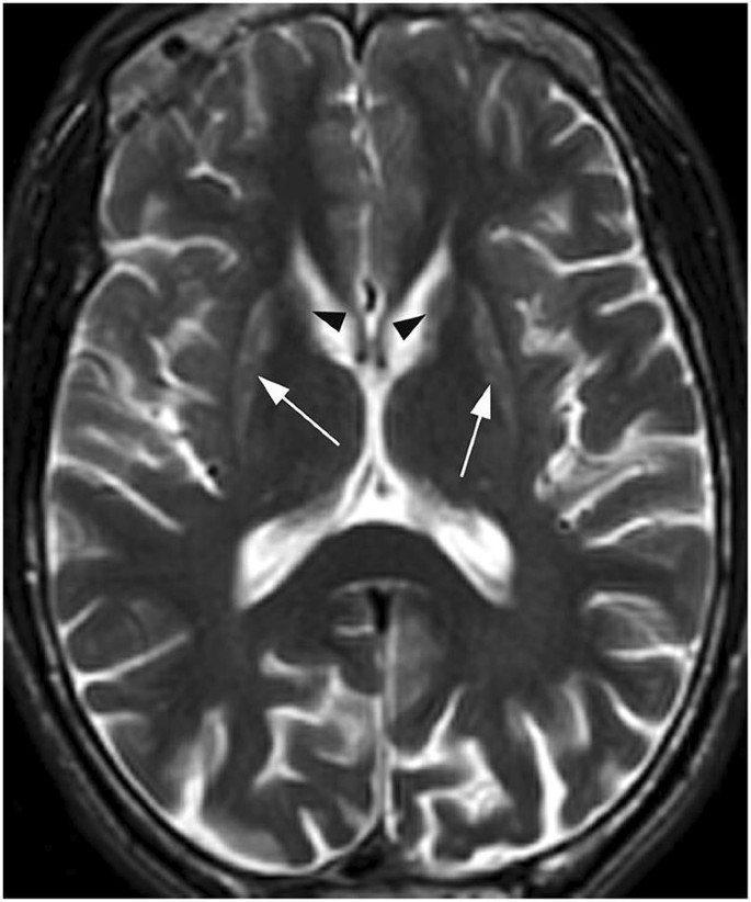
T2-weighted image is showing symmetrical hyperintense signal changes in...  | Download Scientific Diagram
T2-weighted image is showing symmetrical hyperintense signal changes in... | Download Scientific Diagram

T2-Weighted Hyperintensities (Unidentified Bright Objects) in

Bilateral symmetric hyperintensity in globus pallidus on T1-weighted MR  image in a patient with chronic liver disease | Eurorad
Bilateral symmetric hyperintensity in globus pallidus on T1-weighted MR image in a patient with chronic liver disease | Eurorad

Symmetric globus pallidus T2/FLAIR hyperintensity(arrows) (A and B) in... |  Download Scientific Diagram
Symmetric globus pallidus T2/FLAIR hyperintensity(arrows) (A and B) in... | Download Scientific Diagram